Biodegradable MRI imaging agents
Multimeric, biodegradable, imaging agents for MRI that can amongst other things, target lesions in the blood brain barrier.
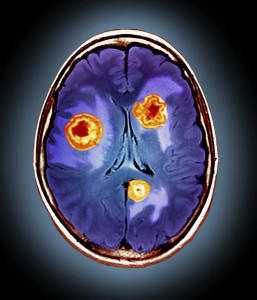
The imaging agents, which are comprised of novel sugar coated iron oxide particles, can be used to identify endothelial cell activation in the brain and in this way the researchers have been able to reveal the presence of pathology that cannot be visualised using conventional imaging techniques.
Data obtained by the researchers demonstrate that these contrast agents could potentially enable early detection of brain injury and inflammation.
The background
Iron-containing particles are known for use in molecular imaging applications, superparamagnetic iron oxide particles (SPIOs) first being employed as magnetic resonance imaging (MRI) contrast agents some years ago. The size and coating of SPIOs helps to determine where the particles end up when administered to a patient, however, it is still difficult to accurately target them to a particular region of interest.
It has been found that small particles (of the order of 50nm to 200nm) provide a strong magnetic resonance signal, while larger particles (around 1μm) provide a much better signal to noise ratio. However, both types of particle suffer from disadvantages. Small particles are often rapidly cleared from the target site, but may still linger in the body. As a result, the quality of repeat MRI scans can be reduced due to the presence of imaging agents from a previous scan. Large particles, on the other hand, can cause microvessel blockage.
There is therefore a need to develop new particles for use as contrast agents that do not suffer from the aforementioned disadvantages.
The Oxford invention
Researchers at the University of Oxford have developed multimeric, biodegradable, imaging agents for MRI that can amongst other things, target lesions in the blood brain barrier. The imaging agents, which are comprised of novel sugar coated iron oxide particles, can be used to identify endothelial cell activation in the brain and in this way the researchers have been able to reveal the presence of pathology that cannot be visualised using conventional imaging techniques. Data obtained by the researchers demonstrate that these contrast agents could potentially enable early detection of brain injury and inflammation.
Commercial opportunity and patent status
The patent is granted in Europe and is still undergoing prosecution in the US. The technology is being progressed under an MRC DPFS project. Oxford University Innovation would be keen to talk to companies interested in developing the commercial opportunities for this technology.
about this technology