Improved scanning probe microscope functionality
Low cost microscope stage with integrated optical light source for use with scanning probe microscopes (SPMs) allowing analysis of drug/target interactions, light-reactive biological and non-biological samples at the molecular level.
Advantages include irradiation levels with low noise and compatibility with almost any SPM.
The invention
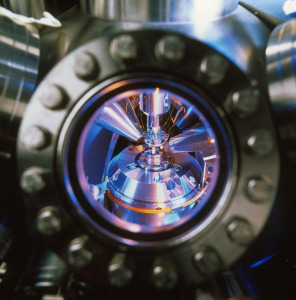
University of Oxford and Nippon Telephone and Telegraph (NTT) researchers have developed a removable microscope stage suitable for use on any Atomic Force Microscope (AFM) to investigate the effects of light from an optical light source on biological or non-biological samples.
Currently there is no commercially available attachment tailored for illuminating AFM samples in this way – previous attempts to meet market need have been unwieldy, SPM-specific and not user-friendly.
The invention can be used to analyse individual receptors in drug-target interactions (see images below: caged drug in solution around a target is released by light and binds the target).
The stage can be tailored to the need, for example, to analyse the effects of UV light or coloured light on biologically active samples, or broad-spectrum light on light-cured resins.
The microscope stage offers many benefits including:
-
low-cost manufacturing process resulting in a big profit margin
-
robust and simple to use on any AFM; many stage designs are envisaged
-
reduced electromagnetic and mechanical noise
-
high level of irradiation at specific wavelengths
Analysis of drug/target interaction:
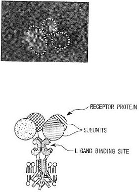
Above: before irradiation (figure 1)
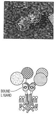
Above: after irradiation (figure 2)
A drug target (‘receptor protein’) is held in solution surrounded by a caged drug (‘ligand’– figure 1). Using the invention, a pulse of light releases the drug that then binds its target, causing the target to change shape (figure 2). Individual target molecules can be examined before and after drug-binding, using an AFM.
Market
This invention is likely to be of interest to scanning probe microscopy manufacturers, businesses engaged in drug discovery or in the development of other light-reactive materials.
The invention is supported by proof-of-concept data, specifically in relation to drug/target interactions.
Patent status
The invention is the subject of a patent application and a scientific publication.
Oxford University Innovation would like to talk to companies interested in developing the commercial opportunity.
Request more information if you would like to discuss this further.
about this technology