Off-resonance Correction Method for Magnetic Resonance Perfusion Imaging and Angiography
Oxford researchers have developed a method for correcting off-resonance effects present in Arterial Spin Labelling (ASL) Magnetic Resonance Imaging (MRI). This increases the effectiveness of non-invasive ASL techniques in the assessment and diagnosis of vascular diseases, such as atherosclerosis and arteriovenous malformation. Improved MRI techniques could remove the need for contrast-enhanced perfusion imaging and X-ray digital subtraction angiography, which requires the insertion of a catheter to administer a contrast agent and the use of ionising radiation. The use of improved ASL methods will greatly increase patient comfort and reduce risks associated with other invasive imaging methods. The Oxford method is simple to implement and allows off-resonance to be corrected without increasing the ASL scan time, removing bias across different vascular territories and boosting signal-to-noise ratio.
Invasive Perfusion Imaging and Angiography – a less than ideal gold standard
Perfusion imaging provides qualitative and quantitative information on blood flow whilst angiographic methods generate images of blood vessels. Both perfusion imaging and angiography are of great importance in the assessment of vascular diseases by providing information on the function and health of tissue and blood vessels in the brain. This knowledge aids clinicians with diagnosis, prognosis and treatment planning in these patients. Most MRI methods for acquiring perfusion information involve administering a gadolinium based contrast agent, which have been linked to nephrogenic systemic fibrosis in patients with kidney dysfunction. Additionally, X-ray digital subtraction angiography is the gold standard for acquiring vessel-specific angiographic information; however, this requires both the insertion of a catheter to administer a contrast agent and the use of ionizing radiation. Associated risks to the patient include strokes or transient ischemic attacks.
Magnetic Resonance Imaging – reduced risk for patients
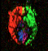
MRI techniques, such as Pseudo-Continuous Arterial Spin Labelling (PCASL) and vessel-encoded PCASL (VEPCASL) are powerful, non-invasive methods available to clinicians to acquire perfusion data and angiograms in the brain without the use of contrast agents. VEPCASL allows acquisition of vascular territory maps and vessel-selective angiograms. VEPCASL has an advantage over other vessel-selective methods in that it allows vessels to be labelled that are closer together. However, off-resonance (magnetic field inhomogeneity) in the labelling plane can occur in either case, leading to a reduction in labelling efficiency and thus image quality. Current methods for off-resonance correction are limited, with some requiring additional PCASL scans and/or manual intervention to calculate the corrections needed.
Key features and commercialisation
- Simple to implement
- Based on generating an Optimised Encoding Scheme (OES)
- Applicable to any pattern of off-resonance and any number of vessels
- Works with both conventional PCASL and VEPCASL
A provisional patent application has been filed in the USA. Oxford University Innovation are seeking partners in the MRI software and /or hardware space to commercialise this technology
about this technology